Dr Ben Wild
Overview |
For vision to be in clear and in focus, light must pass through the cornea, the clear tissue at the front of the eye, the pupil, the black hole in the center of the colored iris, and the lens, the clear tissue inside the eye to the tissue responsible for detecting light, the retina. The lens sits inside a bag (capsule) attached to strings (zonules) that are controlled by muscles (ciliary body).
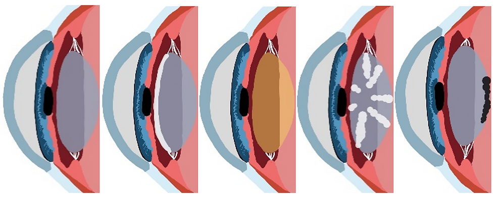
Cataracts refer to the fogging/clouding of the lens. There are many different types of cataracts but the 4 main types are anterior subcapsular cataracts (ASC), nuclear sclerotic cataracts (NSC), cortical cataracts, and posterior subcapsular cataracts (PSC). PSCs tend to be the fastest growing cataract of all.

From left to right, healthy lens, ASC, NSC, cortical cataract and PSC.
Cataracts are most often age related but systemic (full body) diseases, eye diseases, retinal dystrophies, and trauma can lead to early cataracts. Congenital cataracts (cataracts your are born with) can be due to tuberculosis, rubella, shingles, and other infections during pregnancy, metabolic disorders like Fabry disease, galactosemia and more.
Signs and Symptoms |
Signs
Yellow, brown or black lens, white haze or plaque on the lens, bubbles within the lens, or spoke like white haze within the lens.
Symptoms
Constant blurry vision that cannot be corrected with glasses, glare, difficulty with night vision, difficulty with lots of light (PSC), needing more light to see.
Causes and Risk Factors |
Causes
The solidification of crystals within the lens.
Risk Factors
Diabetes, myotonic dystrophy, atopic dermatitis, neurofibromatosis, systemic diseases that cause uveitis (inflammation inside the eye), glaucoma, age, possibly ultra-violet damage, over use of corticosteroids, rubella infection, possibly extended periods of dehydration, high nearsightedness.
Prevention and Treatment |
Prevention
Stay hydrated, wear UV blocking glasses, avoid preventable systemic diseases with a healthy and balanced lifestyle.
Treatments
Cataract surgery when the cataracts start to affect daily activities or when they become too hazy for an eye care professional to properly monitor ocular health.
Surgery starts with topical and systemic anesthesia, although the patient usually remains conscious. 2 incisions are made in the cornea and the eye is filled with viscoelastic gel. A small hole is created in the capsule and fluid inserted to dissect the lens from the capsule. The lens is then cut into pieces and vacuumed out. The capsule is inflated with gel and a plastic intra-ocular lens is inserted into that same capsule. Femtosecond cataract surgery involves using a laser to create the incision, the hole in the capsule and to even break up the natural lens for greater precision.
Intra-ocular Lens Options |
1. Single vision: the surgeon can correct your vision for near or distance but not both.
2. Monovision: Use single vision lenses to correct one eye for distance and one eye for near so that the patient can cope without glasses. This may compromise depth perception.
3. Multifocal: lenses that correct both distance and near prescriptions although fluctuating vision and glare become issues.
Prognosis |
In most cases, a full recovery of vision is expected as long as there are no other ocular diseases present. Most surgeries proceed without any complications and of those rare cases, most complications are easily manageable. These complications may include allergies to eye drops, elevated eye pressures and glaucoma, uveitis (intra-ocular inflammation or infection), and scarring over of the intra-ocular lens (posterior capsular opacification). Other much less-common complications include retinal detachment, double vision, droopy eyelids, leaking of the vitreous gel, breakage of the zonules that hold the capsule, dislocation of the lens, ripping of the capsule, swelling of the retina, fogging of the cornea and risks associated with anesthesia.
