92 items found for ""
- Phlyctenulosis
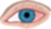
Dr Ben Wild The eyelids are made up of various types of skin, muscle, glands, hair follicles and much more. Their purpose is to protect the eyes from dryness, bright lights and irritants. As with skin everywhere on the body, the eyelid skin harbors bacteria. If the wrong type of bacteria colonize the eyelids or if there is an overgrowth of the normal bacteria, the eyelids can cause harm to the eyes. This harm can involve the cornea, the clear outermost layer of the eye in front of the colored iris, and the conjunctiva, the clear layer over the white part of the eye otherwise know as the sclera. Frontal view of a healthy eye. Phlyctenulosis refers to a hypersensitivity reaction usually within the conjunctiva but sometimes on the edge of the cornea to the bacteria on the eyelids. It is seen as an off-white nodule. If not treated, it can lead to perforation of the eye but usually only lasts 2-3 weeks. If it become chronic or recurrent, it may be associated with a tuberculosis infection. Frontal view of a an eye with a yellow phlyctenule. Signs Off white nodule on the conjunctiva or on the edge of the cornea with redness around the affected area. Symptoms Mild to moderate discomfort, redness, tearing. Causes Allergic hypersensitivity reaction to toxins created by the bacteria on the eyelids. Risk factors Blepharitis, rosacea, eczema, TB infection. Prevention Daily eyelid hygiene. Treatments · Antibiotic eye drops and ointment. · Steroid eye drops. · Oral tetracycline pills. · TB skin test if unresponsive to treatment. Most cases of phlyctenulosis are very easily managed through the aforementioned treatments. Without treatment, a phlyctenule can resolve on its own but are much more likely to cause permanent scarring, ulceration, blood vessel growth into the cornea and in very rare instances, perforation of the eye, which may lead to losing the eye. The causes of these conditions can usually be managed with proper lid hygiene but in some cases, need continuous treatment.
- Recurrent Corneal Erosions
Dr Ben Wild The cornea is the clear tissue in front of the iris (the colored part) of the eye. The cornea is comprised of 5 layers but can be simplified into 3 main layers; the epithelium (outer most layer), the stroma (middle layer) and endothelium (inner most layer). Frontal view of a healthy eye. Recurrent corneal erosions occur when the epithelium dries out at night during sleep, sticks to the back of the eyelids, and upon awakening and opening of the eyelids, rips open. This happens because the epithelium is weakly attached to the underlying cornea. The causes of this poor adhesion are trauma to the eye via infection, scratch, dry eye, or genetic corneal dystrophies such as epithelial membrane dystrophy, Meesman corneal dystrophy, Reis-Buckler’s corneal dystrophy, lattice dystrophy and more. Frontal view of a corneal erosion Signs Red eye, tear in the corneal epithelium, watery eye. Symptoms An immediate stabbing sensation upon opening eyes in the morning, light sensitivity, eye twitch, decreased vision, foreign body sensation. Causes Poor epithelial adhesion to the underlying cornea. Risk factors Dry eyes, trauma, genetic corneal dystrophies. Prevention Avoid exposure to the causes/risk factors above. Treatments · Rolling eyes around underneath lids before opening eyes. · Cyclopentolate drops for pain. · Antibiotic drops. · Bandage contact lens. · Non-steroidal anti-inflammatory drops or pills. · Hypertonic eye drops nightly for 3 months. · Gel drops or ophthalmic ointment. · Surgery: diamond burr polish, stromal puncture, excimer laser. Recurrent corneal erosions can affect quality of life by causing sufferers to become nervous or even scared to open their eyes in the morning. Thankfully, in most situations, recurrent corneal erosions can be easily treated. In some cases, the erosions continue to occur despite best efforts. In these situations, the surgical options can treat 80-90% of cases.
- Ocular Rosacea
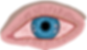
Dr Ben Wild The eyelids are made up of various types of skin, muscle, glands, hair follicles and much more. Their purpose is to protect the eyes from dryness, bright lights and irritants. Frontal view of a healthy eye. Rosacea is a common chronic skin condition that can affect facial skin by causing blood vessels to become visible, blushing, flushing and pustule formation without blackheads, similar to acne. Rosacea is much more common in Caucasians and usually presents along the forehead and both cheeks from the lower eyelid to below the nose or on other sun exposed areas of the skin. Unfortunately, there is no known cure but treatment can reduce or even control the signs and symptoms. Ocular rosacea affects 20% of people with rosacea and presents as dilated blood vessels of the eyelids, pustule formation, and blepharitis. Severe rosacea can affect the eye itself, causing various conditions like phlyctenulosis, marginal keratitis, and the growth of blood vessels into the cornea. Front view of an eye with severe ocular rosacea. Signs Crusty eyelashes, red eyelids, red eyes, loss of eyelashes, recurrent chalazions (eyelid styes), off white bumps on the conjunctiva, collections of white blood cells in the cornea. Symptoms Stinging, burning, gritty, itchy eyes, mild light sensitivity, poor contact lens tolerance (difficulty with contact lens comfort), fluctuating vision. Causes There are no know causes of rosacea but skin becomes irritated after hot drinks, spicy foods, alcohol, extreme temperatures, sun, wind, exercise, and skin care products with preservatives. Risk factors Female, Caucasian, skin that burns easily, smoking, genetic predisposition. Prevention Avoid exposure to the causes/risk factors above. Treatments 1. Artificial tears. 2. Hot compresses and lid massage. 3. Lid hygiene with Blephagel or Cetaphil. 4. Topical antibiotic drops or ointments. 5. Topical steroid drops. 6. Topical cyclosporin drops. 7. Tetracycline pills. 8. Retinoids (although this could make it worse). 9. Intense Pulsed Light (IPL) or radio frequency (RF) treatments. Unfortunately, there is usually no cure for rosacea. It can lead to a lifetime of dry eye symptoms that need regular management. Thankfully, in most cases, regular management including artificial tears, lid hygiene and hot compresses will likely be enough. Severe cases can cause corneal scarring which could affect vision and requires frequent follow up exams and more intensive treatment.
- Pseudoexfoliation Syndrome (Exfoliation Syndrome)
Dr Ben Wild Just like a soccer ball, the eye must stay pressurized to function properly. Normally, the eye is inflated by the production of fluid, called the aqueous humor. This fluid circulates around the inside of the eye providing nutrients and removing wastes created by many eye structures. It exits at the root of the iris, the colored part of the eye, called Schlemm’s canal. Schlemm’s canal is separated from the inner contents of the eye via a fine sieve, called the trabecular meshwork. The interplay between aqueous humor production and the resistance created by the density of the trabecular meshwork creates the pressure inside the eye. Front view (left) and sagittal view (right) of a healthy eye. Pseudoexfoliation syndrome, is a condition whereby fibrous material leaks from the blood vessels of the eye and accumulates on many ocular structures including the conjunctiva, cornea, iris, lens, zonules, and trabecular meshwork. This material can cause early cataracts, can weaken the zonules that hold the lens in place making cataract surgery more difficult, and can clog the trabecular meshwork leading to higher eye pressures and glaucoma in 50-60% of affected individuals. It is rarely noticed before 50 years old. It is now becoming known as exfoliation syndrome because this fibrous material has also been noticed in skin, blood vessels, lungs, and the brain and has been associated with Alzheimer’s disease and hearing loss. Frontal view showing pigment granules on the cornea and exfoliative material on the iris (left) and sagittal view showing the exfoliative material and pigment within the eye (right). Signs White fibrous material on the lens, iris, and trabecular meshwork, loss of iris pigment, pigment granules on the cornea. Symptoms Usually, no symptoms but can cause blurry vision and halos from developing cataracts and progressive blind spots from glaucoma. Causes Fibrous material leaking from blood vessels within the eye or other parts of the body. Risk factors Low levels folate in the blood, female gender, Scandinavian decent. Prevention There is no known way to prevent pseudoexfoliation but treatments can ensure it does not lead to glaucoma. Treatments · Regular glaucoma testing with imaging and peripheral vision testing every 6-12 months. · Glaucoma drops that lower eye pressure. · Laser trabeculoplasty removes the fibrous material in the trabecular meshwork. Exfoliation syndrome itself causes early cataracts and makes cataract surgery more difficult, although still possible. It leads to glaucoma in only about 50% of individuals but this type of glaucoma has a worse prognosis than primary open angle glaucoma because the condition is so variable and difficult to treat. Eye pressures tend to fluctuate dramatically day-day or even within a single day and therefore a treatment may be initiated based on erroneous pressure measurements. Typically though, treatment can ensure glaucoma never advances to a point where the effects can harm day to day life.
- Pinguecula vs Pterygium
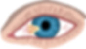
Dr Ben Wild The conjunctiva is a clear layer of tissue that extends from the edge of the cornea, around the visible portion of the eye in front of the white sclera, and even the back surface of the eyelids. It posses as a barrier against foreign material and contributes to the tear film. Frontal view of a healthy eye. A pinguecula and a pterygium are two very similar ocular findings. They both represent degenerations of the conjunctiva leading to the growth of a secondary fibrous, blood vessel rich, layer. Pingueculae are often seen bilaterally (in both eyes) as a yellowish/white mound above the nasal sides of the sclera. Sometimes they can also be found temporally. If severe, they can become inflamed (pingueculitis) or calcified. They never cross into the cornea. Frontal view of an eye with 2 pingueculae. A pterygium is more commonly found on people from countries near the equator. It can be found unilaterally or bilaterally, is most often seen starting from the nasal sides of each eye, but can be found temporally Pterygia look like a wing or triangle of fibrous tissue growing from the conjunctiva over the cornea. Frontal view of an eye with a nasal pterygium. Signs Symptoms Causes Ultra violet radiation and ocular dryness. Risk factors Ultra violet radiation exposure from being outside without UV-blocking glasses or sun glasses. Prevention UV protection and address any dry eye symptoms. Treatments A pinguecula is a very common finding and is considered a normal finding in most cases. In some cases, where a pinguecula has grown substantially, it requires careful management of symptoms. Very rarely does it reach a point where excision is warranted. In some cases where they are surgically removed, they can be recurrent. It is not vision threatening. A pterygium is only vision threatening if it grows far enough into the cornea . Most do not reach this stage. Excision surgery does not usually remove 100% of the tissue and therefore the patient is often left with some irregular astigmatism that cannot be fully corrected with glasses. Sometimes these can recur after surgery.
- Trichiasis vs Distichiasis
Dr Ben Wild Trichiasis and distichiasis are two conditions that result in eyelashes being directed into the eye and causing discomfort. Front view of healthy eyelashes. Trichiasis occurs when the normal eyelashes, which are located on the outside of the eyelid margin, curl inwards towards the eye instead of curling outwards away from the eye. These lashes can scratch the cornea and/or conjunctiva and cause ocular irritation. This can be noticed at birth but is usually acquired. Frontal view of an eye with trichiasis, eyelashes being misdirected towards the eye on both eyelids. Distichiasis occurs when a second row of eyelashes, which are usually much smaller and thinner, grow much closer to the eye right on top of the eyelid margins. They often become misdirected and curl towards the eye. It can present as a full or partial second row of lashes and can be seen right at birth or can be acquired. When seen at birth, distichiasis usually is seen as an entire 2nd row of lashes but usually does not become irritating until around 5 years old. Acquired distichiasis is usually seen as only a few extra slow growing lashes, however, are usually more symptomatic. Frontal view of an eye with distichiasis, a second row of lashes on both eyelids. Signs Symptoms Both: Irritation worse when blinking. Both: Inflammation from blepharitis, shingles, trauma, eyelid surgery, thermal/chemical burns, Stevens-Johnson syndrome, ocular cicatricial pemphigoid and more auto-immune related skin conditions. Prevention Control inflammation. Treatments · Epilation (tweezing lashes every 1-2 months). · Electrolysis of the lash follicle. · Laser ablation of the lash follicle. · Cryotherapy of the lash follicle. Epilation is the easy fix, but more often than not, the lashes regrow. Electrolysis, laser hair removal or cryotherapy usually are more permanent solutions. If this condition is treated properly, there should be no permanent vision loss.
- Pigment Dispersion Syndrome
Dr Ben Wild Just like a soccer ball, the eye must stay pressurized to function properly. Normally, the eye is inflated by the production of fluid, called the aqueous humor. This fluid circulates around the inside of the eye providing nutrients and removing wastes created by many eye structures. It exits at the root of the iris, the colored part of the eye, called Schlemm’s canal. Schlemm’s canal is separated from the inner contents of the eye via a fine sieve, called the trabecular meshwork. The interplay between aqeous humor production and the resistance created by the density of the trabecular meshwork creates the pressure inside the eye. Front view (left) and sagittal view (right) of a healthy eye. Pigment dispersion syndrome is typically a bilateral (both eyes) condition whereby the iris develops in such a way that it bows backwards and rubs against the zonules (strings that hold the lens of the eye in place). This continual rubbing results in the pigment granules, that give the iris its color, becoming liberated from the iris. These floating granules follow the same flow as the aqueous humor and start to accumulate on the lens of the eye, the cornea, and most importantly, the trabecular meshwork. The trabecular meshwork then becomes blocked with pigment and creates even more resistance for aqueous humor outflow leading to elevated eye pressures. These elevated pressures can lead to glaucoma in about 35% of affected individuals. Frontal view showing pigment granules on the cornea (left) and sagittal view showing the iris bowing and pigment granules on the cornea, lens, trabecular meshwork and retina Signs Pigment granules stuck to the inside of the cornea, granules stuck to the lens, granules floating in the eye, granules depositing on the retina, elevated eye pressures, and the loss of iris pigment where the zonules are located. Symptoms Usually no symptoms but sometimes, if severe, can lead to fast onset of blurring vision, headache, and glare often after exercising. Causes Abnormal development of the eye leading to the iris bowing backwards and rubbing on the zonules. Risk Factors High myopia (nearsightedness), male gender, Caucasian ethnicity. Prevention There is no known way to prevent pigment dispersion syndrome but treatments can ensure it does not lead to glaucoma. Treatments · Regular glaucoma testing with imaging and peripheral vision testing. · Laser peripheral iridotomy to stop the iris from bowing backwards. · Pilocarpine drops before exercising, this shrinks your pupil to straighten out the iris. · Laser trabeculoplasty to remove the pigment in the trabecular meshwork. Pigment dispersion syndrome itself is not a condition that will affect vision but it does put the individual at a higher risk of developing glaucoma, a condition that can lead to the development of large, irreversible, blind spots. Early detection and proper management can ensure that this condition will not affect one’s vision.
- Scleral Hyaline Plaque
Dr Ben Wild The sclera is the white, durable, layer of the eye. Its function is as a strong barrier that holds the contents of the eye. It extends from the cornea at the front of the eye to the optic nerve at the back of the eye. It also acts as an attachment scaffold for the muscles of the eye (extraocular muscles). Frontal view of a healthy eye. Scleral hyaline plaque and senile scleromalacia refer to the thinning of the sclera around where the eye muscles attach to the eye. This is a normal finding that becomes more common with age. Scleral hyaline plaques slowly progress over time whereas senile scleromalacia often sudden occurs. Both of these findings are purely cosmetic and do not affect the eye. Front view of an eye with scleral hyaline plaque/senile scleromalacia. Signs Brown/blue, oval, sharply demarcated areas of thinning of the sclera. Symptoms No symptoms. Causes Tensile pulling forces on the sclera by the extraocular muscles. Risk factors Age. Prevention There are no known preventative measures. Treatments No treatment required. Any concerns about scleral hyaline plaque/senile scleromalacia are purely cosmetic. They do not lead to perforation of the eye or affect vision in any way.
- Episcleritis
Dr Ben Wild The conjunctiva is a clear layer of tissue that extends from the edge of the cornea, around the visible portion of the eye, in front of the white sclera, and even the back surface of the eyelids. It posses as a barrier against foreign material contributes to the tear film. The episclera is a second clear layer sandwiched between the conjunctiva and the white sclera. It’s function is to encapsulate the eye and help protect the contents of the orbits. Frontal view of a healthy eye. Episcleritis is a common condition, usually without a known cause, often occurring in both eyes, involving inflammation of the episclera. This condition can be recurrent and, if so, is more likely to be associated with known conditions. Unlike conjunctivitis, where the blood vessels are straight or wavy, episcleral vessels are radial. There are two main types of episcleritis. Simple episcleritis is 75% of cases and usually peaks at the 24-hour mark after it has been first noticed, then fades. It presents as sectoral (small patch) or diffuse (all-over) redness of the eye. Nodular episcleritis develops very slowly and takes much longer to resolve. It often presents as pain upon awakening, redness worsens and becomes uncomfortable when it develops into a nodule. Front view of an eye with nodular episcleritis on the left of the picture and simple sectoral episcleritis on the right. Signs Symptoms Causes Inflammation of the episclera. Risk factors Dry eyes, rosacea, contact lens wear, collagen vascular diseases like rheumatoid arthritis, shingles. Prevention There are no known preventative measures. Treatments · Most episodes do not need treatment. · Artificial tears. · Cold compresses. · Non-steroidal anti-inflammatory pill or drop. · Steroid drop. Episcleritis is not a condition that can affect long term vision or eye health. Although, depending on the cause, it could spread to a different tissue type that could affect the health of the eye, like scleritis. Recurrent episodes can lead to the blood vessels of the eye becoming permanently enlarged.
- Giant Papillary Conjunctivitis
Dr Ben Wild The eyelids are made up of various types of skin, muscle, glands, hair follicles and much more. Their purpose is to protect the eyes from dryness, bright lights and irritants. Frontal view of a healthy eye with the upper lid flipped upwards. Giant papillary conjunctivitis (GPC) occurs due to mechanical rubbing of foreign bodies on the inside of the upper lids. This rubbing causes a large allergic response where the inside of the upper eyelid develops large papillae (bumps containing immune response cells) and mucous secretions. Frontal view of an eye with the top eyelid flipped upwards exposing GPC. Most often, GPC occurs from accumulation of protein deposits on monthly disposable or biweekly disposable contact lenses that rub the eyelid each time a patient blinks. Alternatively, it can be caused by ocular prostheses (either cracked or poorly maintained), sutures after ocular surgery, scleral buckles after a retinal tear or detachment, filtering blebs after glaucoma surgery or even in cases of vernal keratoconjunctivitis or atopic keratoconjunctivitis (both types of allergic conjunctivitis). Signs Large papillae (bumps of immune cells) on the inside of the upper eyelids, mucous discharge. Symptoms Ocular irritation, contact lens intolerance, foreign body sensation, itchiness. Foreign bodies (protein on biweekly or monthly disposable contacts, rigid contact lenses, stitches, prosthetics, scleral buckles, blebs, etc.), severe allergies. Prevention Start with daily disposable contacts, avoid wearing contact lenses overnight, avoid wear contact lenses over 14 hours a day (different number for everyone), daily cleaning of ocular prosthetics. Treatments · Mast cell stabilizer drops. · Antihistamine drops. · Dual action antihistamine/mast cell drops. · Steroid drops. · Non-steroidal anti-inflammatory drops. · Discontinue contact lens wear for several weeks. · Decrease contact lens wear time. · Change contact lens storage solution. · Switch to daily disposable contacts. · Laser eye surgery to avoid contact lens wear. · Removal of scleral buckle. · Removal of exposed sutures. GPC is not a vision threatening condition but it can cause substantial ocular discomfort and contact lens intolerance (contact lenses become too uncomfortable to wear). It is also very difficult to treat and sometimes, although rarely, result in permanent contact lens intolerance. To avoid this fate, the treatments above should be started as soon as symptoms develop.
- Horner Syndrome
Dr Ben Wild The iris, the colored part of our eyes, contains 2 muscles. One, the sphincter, is controlled by the parasympathetic nervous system and the other, the dilator, is controlled by the sympathetic nervous system. The interplay between these muscles controls the size of the opening in the middle of the iris known as the pupil. A frontal view of a healthy eye. Horner syndrome is a condition that is usually unilateral whereby the sympathetic nervous system pathway is disrupted. In rare cases it may be bilateral when caused by issues like diabetes. There is a 3-link chain in this sympathetic nervous system pathway and if either of these links are severed, it results in a small pupil, a droopy eyelid, and the loss of the ability to sweat on that side of the face. One can be born with this condition. A frontal view of an eye with Horner Syndrome showing a small pupil even in dim light and a droopy eyelid. The first link is a neuron connecting the hypothalamus (brain) to the spinal cord. Issues such as diabetes, stroke, brain tumors, spinal cord tumors, Multiple Sclerosis, and more can cause this. The second goes from the spinal cord to the cervical ganglia located just outside the spinal cord. Issues like apical lung tumors aortic or carotid aneurysms and neck trauma can cause this. The third goes from the cervical ganglia up the internal carotid artery and into the eye. Issues like trauma, pituitary tumors, carotid artery issues, jugular vein issues, aneurysms, and inflammation can cause this. Signs Different sized pupils (unless bilateral), droopy eyelid, loss of the ability to sweat on the affect side of the head, lighter iris if born with the condition. Symptoms Pain if due to a cause located in the neck (carotid or jugular issues). Causes Loss of sympathetic nerve supply to the affect eye. Risk Factors Brain tumors, brain aneurysms, spinal cord tumors, carotid aneurysms, jugular vein issues, inflammation, diabetes, trauma, MS, strokes, lung tumors, surgery. Prevention Regular check ups with a family doctor to catch any health conditions early and live a healthy lifestyle to avoid vascular conditions like diabetes. Treatments commence after determining the cause of the Horner’s pupil via CT/MRI scans. · Surgery to remove the lesion. · Apraclonidine drops to dilate the affected pupil (depends on which neuron is not working). Horner syndrome can be a sign of a life-threatening condition and must be taken seriously. If it presents acutely (suddenly), it necessitates an immediate visit to the emergency room. If it has been slowly appearing over the course of a year, it still necessitates investigation but is not an emergency. The eye symptoms are not typically bothersome and do not require treatment.
- Filamentary Keratitis
Dr Ben Wild Dry eye represents a breakdown of the complex interplay between tear volume, tear film stability (evaporation), inflammation, and eyelid function. The lacrimal gland produces most of the watery (aqueous) portion of the tears responsible for oxygen transmission, removal of debris, and antimicrobial activity. The meibomian glands, located along the edges of the eyelids, produce the oil (lipid) portion of the tears responsible for protecting the aqueous portion from evaporation. Lastly, there is a mucous portion, found attached to the cornea and conjunctiva that helps in spreading the tears for lubrication. Frontal view of a healthy cornea. Filamentary keratitis refers to a common, advanced state, of dry eye whereby strands of epithelium, the outermost layer of the cornea, mucous, and cell debris form on the cornea. These strands tend to be strongly attached to the cornea. With each blink, the strands are pulled in one direction or another and cause considerable discomfort. Frontal view of a cornea with filaments. Signs Red eyes, stringy discharge, low tear volume, filaments attached to the cornea. Symptoms Foreign body sensation, sometimes light sensitivity, dry eyes. Causes Extreme dry eye from exposure of the eye, low tear volume, poor tear composition. Risk Factors Lagophthalmos (eyelids never shutting completely), autoimmune conditions (Rheumatoid arthritis, Sjogren’s, etc.), medications causing dry eyes, excessive contact lens use, cataract surgery, laser eye surgery, causes of neurotrophic keratitis. Prevention Supplement your tears with artificial tears if you are taking a medication or have an eye condition listed above. Maintain proper lid hygiene, maintain a diet rich in omega 3s, avoid smoking, avoid dehydration, take 20 second breaks every 20 minutes to blink when reading or on the computer. Treatments (depending on the cause) · Preservative free artificial tears. · Alter medications. · Remove filaments one by one. · Hypertonic (salt) eye drops. · Bandage contact lens to help with the pain. · Mucolytic agents to break up the mucous strands. · Cyclosporin drops for pain. · Steroid drops to decrease inflammation. · Punctal plugs to allow tears to stay on the eyes longer. Filamentary keratitis, in most cases, is very manageable. With that being said, depending on the cause, there may be no cure. In most cases, filamentary keratitis will be only a minor inconvenience of comfort but in severe cases, can cause a lifetime of extreme discomfort, vision fluctuations, and ocular surface diseases.